Anatomy and physiology of the eyelids

Anatomy and physiology of the eyelids
 the eyelids are vital to the maintenance of ocular surface integrity.
mechanical barrier to a variety of insults
sweeping mechanism to remove debris from the cornea (blink reflex)
vital contribution to the production and drainage of the tear film.
contribute to facial expression, and even minor aberrations or asymmetry may affect cosmesis.
the lids comprise a layered structure of
• skin
• orbicularis oculi
• tarsal plates/septum
• conjunctiva
the orbital portion is more complex, with preaponeurotic fat and retractors lying deep to the septum.
the interpalpebral fissure is usually 30mm wide and 10mm high (slightly higher in  ).
).
the resting position of the upper lid is 2mm below the superior limbus (higher in children)
the lower lid, the resting
Skin and eyelashes in Anatomy and physiology of the eyelids
the skin of eyelids is very thin and has loose connective tissue but no subcutaneous fat.
contains eccrine sweat glands and sebaceous glands.
the lashes
• arranged in 2–3 rows along the lid margins
• about 150 on the upper and 75 on the lower lid.
• replaced every 4–6 months
• grow back faster if cut.
• the lash follicles have apocrine sweat glands (of Moll) and modified sebaceous glands (of Zeis).
 Orbicularis oculi in Anatomy and physiology of the eyelids
thin sheet of striated muscle is divided into orbital and palpebral portions
palpebral portion is further divided into preseptal and pretarsal parts.
Innervated by temporal and zygomatic branches of VIIn for the orbicularis overlying the upper lid and by the zygomatic branch alone for the lower lid.
the orbital portion forms a ring of muscle arising from the medial canthal tendon and parts of the orbital rim.
the preseptal part of each lid runs from the medial canthal tendon and arches over the anterior surface of the orbital septum, and inserts into the lateral horizontal raphe.
each pretarsal part arises from the medial canthal tendon and arches over the tarsal plates and inserts into the lateral canthal tendon and horizontal raphe.
horner muscle is formed by deep pretarsal if bres running medially to insert on to the lacrimal crest.
Functions of the orbicularis oculi include lid closure and the lacrimal pump mechanism.
Orbital septum and tarsal plates in Anatomy and physiology of the eyelids
the septum is a sheet of tissue that arises from the orbital rim where it is continuous with orbital fascia and periosteum.
towards the palpebral margin, Orbital septum is thickened, forming the tarsal plates that maintain the shape of the lid.
Tarsus 25mm long, 1 mm thick, and of variable height 10mm high for the upper lid and 5mm for the lower lid.
they also contain MGs (about 35 in the upper lid, 25 in the lower lid) which secrete the lipid component of the tear film.
Canthal tendons in Anatomy and physiology of the eyelids
At each end, the tarsal plates are stabilized by a horizontal canthal tendon.
the medial canthal tendon is well developed with an anterior limb arising from the anterior lacrimal crest, and a posterior limb from the posterior lacrimal crest.
the lateral canthal tendon lies just posterior to the horizontal raphe and inserts into the zygomatic bone (Whitnall’s tubercle) and merges posteriorly with the lateral check ligament (from the sheath of lateral rectus).
Fat pads in Anatomy and physiology of the eyelids
The preaponeurotic fat pads are extensions of orbital fat lying just posterior to the orbital septum.
Lid retractors in Anatomy and physiology of the eyelids
the upper lid retractors comprise levator palpebrae superioris (LPS) and Müller’s muscle.
LPS originates from the orbital apex and runs forward over superior rectus (SR) to the orbital rim.
At this point, it is stabilized by the superior transverse ligament of Whitnall (a fascial bridge running between the trochlea and the lacrimal gland fascia) permitting the distal LPS to run steeply downward and insert as an aponeurosis into the septum, tarsus, and orbicularis.
LPS Innervated by IIIn and Müller’s muscle is an accessory retractor muscle supplied by the sympathetic system.
Müller muscle overaction is demonstrated in sympathetic overdrive and TED and underaction is seen in horner’s syndrome.
the lower lid retractors are more rudimentary but are similarly divided into voluntary and sympathetic groups.
The lower eyelid retractor
• fascial extension from the terminal muscle fibers and tendon of the inferior rectus muscle, originating as the capsulopalpebral head.
• As it passes anteriorly from its origin, it splits to envelop the inferior oblique muscle and reunites as the inferior transverse ligament (Lockwood ligament).
• From there, the fascial tissue passes anterosuperiorly as the capsulopalpebral fascia.
• The bulk of the capsulopalpebral fascia inserts on the inferior border of the inferior tarsus.
• Fibers also pass forward, to the inferior fornix conjunctiva and through orbital fat to the orbital septum, and forward to the subcutaneous tissues forming the lower eyelid crease.
• The orbital septum fuses with the capsulopalpebral fascia approximately 5 mm below the inferior tarsal border.
• The inferior tarsal muscle (Müller muscle) lies just posterior to the fascia and is intimate with its structure.
• The sympathetically innervated smooth muscle fibers are first noted near the origin of the capsulopalpebral head.
• The capsulopalpebral head splits into 2 portions to pass around the inferior oblique muscle sheath; the portion beneath the muscle is thin and devoid of smooth muscle, while the portion above is a much thicker fascial layer and contains the smooth muscle fibers. As they continue to pass forward, the smooth muscle fibers do not insert directly onto the inferior tarsal border but into the fascia several millimeters below the tarsal border.
• In the Asian lower lid, the line of fusion of the orbital septum to the capsulopalpebral fascia is often higher, or indistinct, with anterior and superior orbital fat projection, and overriding of the preseptal orbicularis oculi over the pretarsal orbicularis.
• Lower Lid Retractors are of outmost important for lower lid surgeries or even surgeries on IO and IR muscles
Conjunctiva in Anatomy and physiology of the eyelids
conjunctiva is a mucous membrane comprising non-keratinized epithelium, BM, and stroma.
the epithelium of the palpebral conjunctiva is of stratified squamous form.
It contains mucin-secreting goblet cells and crypts of henle.
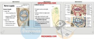
Nerves in Anatomy and physiology of the eyelids
Sensation to the lower lid is mainly by the infraorbital nerve (Vb), with infratrochlear branch of the nasociliary nerve (Va) innervating the medial canthal area.
Sensation to the upper lid is by lacrimal, supraorbital, and supratrochlear nerve (all Va).
orbicularis oculi is innervated by VIIn
LPS by IIIn, and Müller’s muscle by the sympathetic system.
lower lid Retractors by IIIn and sympathetic system
Arteries in Anatomy and physiology of the eyelids
Arterial supply is by three arcades that form anastomoses between the medial palpebral artery (from the terminal ophthalmic artery) and the lateral palpebral artery (from the lacrimal artery).
In the upper lid, there is a marginal arcade 2mm above the margin and a peripheral arcade at the top of the tarsal plate.
In the lower lid, the arcade lies 4mm below the margin.
Veins in Anatomy and physiology of the eyelids
to superficial temporal vein laterally
to the ophthalmic and angular veins medially.
Lymphatics in Anatomy and physiology of the eyelids
to the parotid glands laterally
to the submandibular glands inferiorly
to the anterior cervical chain inferomedially.
Anatomy and physiology of the eyelids powerpoint presentation :
Lecture outline Eyelid anatomy • Gross anatomy • Layers of the eyelid • Eyelid arterial supply and venus and lymphatic drainage • Eyelid nerve supply Eyelid physiology • Functions of eyelids • eyelid movements: Openning,clouser,bli nking and winking,bell’s -Dynamics of eyelid openning and clouser
Anatomy and physiology of the eyelids videos :
Eyelid Anatomy
Anatomy and physiology of the eyelids

 留言列表
留言列表