PIXNET Logo登入

金台診所醫學美容的部落格

跳到主文

歡迎光臨金台診所附設醫學美容在痞客邦的小天地 金台診所醫學美容成立以來.感謝大家的支持與肯定.借此園地與醫美感興趣者相互溝通及交流.不吝賜教.感激萬分. 住址:新台北市三重區市中正北路346號2樓.(02-29844451.29833896) 有醫學美容(雷射光療.微整型(類肉毒Botox.玻尿酸.微晶瓷.膠原蛋白等)及雙眼皮手術需求者 請來電咨詢:02-29844451. 01| 專長 內外科,眼科,耳鼻喉科,家醫科,皮泌科,婦科,專門眼部整形手術,過敏性結膜炎,過敏性鼻炎,氣喘,一位性皮膚炎,塵蜹檢測及減敏治療,健康減重門診治療,醫學美容諮詢,附設:視力保健,隱形眼鏡特別門診 02| 主治科別 塵蜹檢測及減敏治療,健康減重門診治療,醫學美容諮詢 03| 其他科別 內科,外科,眼科,耳鼻喉科,家庭醫學科,皮膚科,泌尿科,婦產科,美容醫學科,專門眼部整形手術

部落格全站分類:醫療保健

  • 相簿
  • 部落格
  • 留言
  • 名片
  • 7月 30 週二 201918:32
  • (新)眼皮 割雙眼皮 眼皮整形 縫雙眼皮 雙眼皮手術 公告:請先矯正屈光不正.再進行雙眼皮手術.萬分感激.

anatomy-physiology-of-eyelids-15-638.jpg
公告:請先 矯正屈光不正 .再進行雙眼皮手術.萬分感激. 
 
(繼續閱讀...)
文章標籤

金台診所附設醫美 發表在 痞客邦 留言(0) 人氣(2,847)

  • 個人分類:醫學美容
▲top
  • 6月 22 週一 202009:01
  • Summary of VF defects


Summary of VF defects


Dr.Reda Gomah El GariabyDr.Reda Gomah El Garia

August 17, 2018

inOphthalmology


182 8

0


(繼續閱讀...)
文章標籤

金台診所附設醫美 發表在 痞客邦 留言(0) 人氣(43)

  • 個人分類:醫學美容
▲top
  • 4月 10 週五 202009:59
  • 什麼是黃斑部病變? 文自康健知識庫


什麼是黃斑部病變?
 
人體視覺最敏感處就是黃斑部,它位在眼球正後方的視網膜中心處。此病是好發於老年人的慢性疾病,造成50歲以上老年人視覺嚴重損害的首要原因。患者會逐漸發生中央黃斑部的退化現象,進而影響患者中央視覺,造成患者視野出現視物扭曲、變形,沒有妥善治療,最終有可能導致法定失明的情況。
 
(繼續閱讀...)
文章標籤

金台診所附設醫美 發表在 痞客邦 留言(0) 人氣(9)

  • 個人分類:
▲top
  • 4月 09 週四 202010:41
  • Coronavirus disease 2019


COVID-19
(Coronavirus disease 2019)


Abstract: COVID-19, also known as Coronavirus disease, is a respiratory infection caused by severe acute respiratory syndrome coronavirus 2 (SARS-CoV 2). Transmission mainly occurs via direct contact or aerosol droplets. The infection may present asymptomatically or with fever and dry cough. Individuals who are over 65 years of age, immunosuppressed, or have preexisting conditions have a higher risk of developing severe symptoms and complications. Management is based on supportive care. 


Updated April 7, 2020


Coronaviruses viewed under an electron microscope. Note the characteristic crown-like (corona) appearance
Image: by CDC/ Dr. Fred Murphy, License: Public Domain Files


Table of Contents


  •  


  •  


  •  


  •  


  •  


  •  


  •  


  •  


  •  


  •  


  •  


  •  


  •  


  •  


Etiology


Coronaviruses (CoV) are a family of enveloped, positive-sense, single-stranded RNA (+ssRNA) viruses. They tend to cause mild upper respiratory diseases in humans. Of the 7 known species of CoV, only 3 are known to cause severe infections in humans:


  • Severe acute respiratory disease coronavirus (SARS-CoV): emerged in 2003 in southern China from civet cats

  • Middle East respiratory syndrome coronavirus (MERS-CoV): emerged in 2012 in Saudi Arabia from dromedary camels

  • SARS-CoV 2: emerged in December 2019 in China possibly from bats or pangolins (still under investigation)


Selected diseases caused by Coronaviruses


 Common coldGI tract infectionSevere acute respiratory syndrome (SARS)COVID-19 (Wuhan City, China)
Incubation3 days3 days4–6 days2–14 days
IncidenceMost commonRareRare~ 1,200,000 cases (Dec 2019 – April 2020)
PrognosisComplete resolutionComplete resolution
(up to 25% fatal for NEC)
30% resolution
70% severe infection
10% fatal
80% resolution
15% severe infection
5% critical infection
5.6% fatal (based on confirmed cases as of April 7, 2020, may change)
Clinical manifestationSneezing, rhinorrhea, headache, sore throat, malaise, fever, chillsDiarrhea, gastroenteritis, neonatal necrotizing enterocolitis

Fever > 37,8°C (100,0°F), muscle pain, lethargy, cough, sore throat, malaise


Shortness of breath/ pneumonia (direct viral or secondary bacterial)


Asymptomatic 


Mild infection: fever, dry cough, malaise, dehydration


Severe infection: high fever, shortness of breath, chest pain, hemoptysis


Complications: pneumonia, ARDS, sepsis, multi-organ failure


NEC: Necrotizing enterocolitis ARDS: Acute respiratory distress syndrome


The SARS-CoV 2 virion is ~1,250 nm in diameter, and its genome ranges from 26 to 32 kilobases, the largest for an RNA virus. It has 4 structural proteins: spike (S), envelope (E), membrane (M), and nucleocapsid (N). 


  • N protein forms a complex with RNA and aids in the viral assembly after its replication.

  • S, E, and M proteins create the viral envelope. 

  • S protein, is a club-shaped surface projection, giving the virus its characteristic crown-like appearance on electron microscopy. It’s responsible for viral entry into the human cell.


Structural proteins of the SARS-CoV 2 virion.
Image: by Lecturio


SARS-CoV 2 attaches to the host cell by binding its S proteins to the receptor protein, angiotensin-converting enzyme 2 (ACE2). ACE2 is expressed by epithelial cells of the intestine, kidney, blood vessels, and most abundantly in type II alveolar cells of the lungs. The virus induces a drop of ACE2 in human cells, possibly inducing lung damage.


The human enzyme transmembrane protease, serine 2 (TMPRSS2) is also used by the virus for S protein priming and to aid in membrane fusion.


Related Video Courses:


Coronavirus Infectious Disease 2019 (COVID-19)


COVID-19 Etiology, Epidemiology, Diagnosis


Transmission


CoV are zoonotic or transmitted to humans through animals. It is hypothesized that horseshoe bats are the natural reservoir of SARS-CoV 2 since its genome is 97% identical to that of a bat coronavirus. The intermediate host is still unknown.


Once in humans, the virus is transmitted mainly via inhalation of aerosol droplets from coughing, sneezing, or talking of symptomatic individuals. In the air, larger droplets tend to drop towards the ground within 1 m (3 ft), while smaller droplets can travel as an aerosol cloud over 2 m (6 ft) and remain viable in the air for up to 3 hours under certain conditions. Other forms of transmission include:


  • Direct transmission through hand-to-face contact from infected surfaces

  • Fecal-oral transmission is hypothesized (observed in SARS infection, but is still under investigation)

  • Vertical transmission (mother-to-child) hasn’t been reported


The period of highest infectivity for symptomatic cases ranges from 2 days before the onset of symptoms up to 3 days after their resolution. (exact limits are still under investigation).


The reproductive number (R0), or the number of secondary infections generated from 1 infected individual, is 2 to 2.5, higher than for influenza (0.9-2.1). COVID-19 is highly contagious due to the following aspects: 


  • Asymptomatic individuals are also infectious, albeit to a slightly lesser degree, posing a real challenge for contagion prevention. 

  • Production of high viral loads

  • Efficient and prolonged shedding of virions from the upper respiratory tract (URT)

    • Median duration of viral RNA shedding from URT: 20 days


  • Can remain infectious on surfaces outside a host from a few hours up to a few days.

    • Viral life-span depends on the type of surface, temperature, and humidity levels

    • There is currently no evidence to suggest that COVID-19 can be acquired from mail and packaged goods.


Image: by Lecturio, Source: The Journal of Hospital Infection


Epidemiology


The first case of COVID-19 was traced back to Wuhan, China, in late November 2019, with an outbreak developing in December. The virus quickly spread, with widespread ongoing transmission occurring globally. More than 1 million people were infected and over 55,000 died within the first 4 months of global spread. COVID-19 was characterized as a pandemic on March 11, 2020.


Coronavirus COVID-19 Global Cases by Johns Hopkins CSSE. https://www.arcgis.com/apps/opsdashboard/index.html#/bda7594740fd40299423467b48e9ecf6. 


Clinical Presentation


The incubation period for COVID-19 ranges from 2–14 days, with an average of 5 days. 

  • 80% of infections are mild or asymptomatic

  • 15% are severe infections (requiring oxygen therapy)

  • 5% are critical infections (requiring ventilation) 


This proportion of severe and critical to mild cases is higher than in influenza infections.


Asymptomatic cases:


  • Can transmit the virus

  • Represent >50% of all infections (still under investigation)

  • May not develop any noticeable symptoms

  • Anosmia, hyposmia, and dysgeusia have been reported in many laboratory-confirmed cases of patients that were otherwise asymptomatic.

  • It has not been clearly determined how long asymptomatic individuals remain contagious after initial infection.


Mild cases:


  • Dry cough and moderate fever

  • Common flu-like symptoms, including fatigue, malaise, runny nose, nasal congestion, and sore throat

  • Less frequently: diarrhea, nausea, vomiting, diffuse abdominal pain, productive cough, headache, and muscle or joint pain

  • Recovery time: ~2 weeks


Clinical presentation of COVID-19.
Image: by Lecturio


Severe cases and complications:


  • ~1 in 6 people with COVID-19 experience clinical deterioration and/or develop a complication in the 2nd week of illness

  • Median time from onset of symptoms to the onset of critical care/ICU transfer: 8-9 days 

  • Patients develop dyspnea, high fever, chest pain, hemoptysis, respiratory crackles, and progressive respiratory failure 

  • Recovery time: ~3-6 weeks


Common complications of COVID-19: viral/interstitial pneumonia, acute respiratory distress syndrome (ARDS), sepsis, and septic shock. Other complications: cardiac injury, arrhythmia, liver dysfunction, acute kidney injury, and multi-organ failure.


Risk factors for the severe form and complications of COVID-19:


  • Immunosuppression (from long-term steroid use, cancer, AIDS/HIV infection, congenital immunodeficiency, use of immunosuppressants, etc.)

  • Age > 65 years 

  • Chronic diseases (especially hypertension, diabetes mellitus, coronary heart disease, and cerebrovascular disease)

  • Pregnancy


The expression of ACE2 is highly increased in patients being treated with ACE inhibitors or angiotensin II type 1 receptor blockers (ARBs). Contrary to initial reports, the American College of Cardiology has pointed out that there is no data to support the claim that ACE inhibitors and ARBs increase the risk of COVID-19 infection, and recommends that patients who already use these medications should continue to do so while further studies are performed.


Refractory cases: 


Nearly 50% of COVID-19 patients did not achieve clinical and radiological remission within 10 days of hospitalization. Patients with male sex, older age, anorexia, and no/low fever at the time of admission have a higher risk of presenting a refractory progression.


Related Video Courses:


Pneumonia (Including Interstitial)


Acute Respiratory Distress Syndrome (ARDS)


Severe Sepsis and Septic Shock


Pulmonary Radiology


Diagnostics


Polymerase chain reaction (PCR) is currently the only test being used to confirm cases of COVID-19 infection and should be performed as soon as possible once a person under investigation (PUI) is identified. The specimens used for testing include: 


  • Nasopharyngeal (NP) and/or oropharyngeal (OP) swab (for mild or asymptomatic suspected cases)

    • NP is the first choice. OP swabs are acceptable only if other swabs are not available.

    • Can be negative initially. If suspicion of COVID-19 remains, retest every 2-3 days. 

    • In severe cases, NP and OP swabs may be negative, while specimens from the lower respiratory tract are positive. 


  • Sputum (for patients with productive cough, inducing is not recommended)

  • Bronchial and tracheal secretions or bronchoalveolar lavage (for patients receiving invasive mechanical ventilation)


Image: by Lecturio


1. Nasopharyngeal swab: Insert swab into a nostril parallel to the palate, and carefully slide it forward until a soft resistance is felt. Swab should reach a depth equal to distance from nostrils to outer opening of the ear. Rotate for several seconds to absorb secretions, and then slowly remove. 1. Oropharyngeal swab: Insert swab into the oral cavity without touching the gums, teeth, and tongue. A tongue depressor may be used. Swab the posterior pharyngeal wall using a rotatory motion. 2. Place swabs immediately into sterile tubes containing 2-3 ml of viral transport media. If both swabs are collected, they should be combined into a single vial. 3. Carefully leverage the swab against the tube rim to break the shaft at the scoreline. 4. Store specimens at 2-8°C for up to 72 hours after collection. If a delay in testing/shipping is expected, store specimens at -70°C or below. Use only synthetic fiber swabs with plastic shafts. Calcium alginate swabs or swabs with wooden shafts may inactivate the virus and inhibit PCR testing. 


During an ongoing COVID-19 outbreak, laboratory testing should be prioritized as follows according to the CDC:


  1. Hospitalized patients with compatible signs and symptoms (especially those presenting with unexplained viral pneumonia or respiratory failure)

  2. Healthcare professionals with compatible signs and symptoms

  3. Symptomatic individuals who are at high risk of developing a severe form of the disease or a complication (e.g., patients who are elderly, immunocompromised, or have chronic conditions) 

  4. Critical infrastructure workers with compatible signs and symptoms

  5. Any individual, including healthcare professionals, who had close contact with a suspect or laboratory-confirmed COVID-19 patient within 2 weeks of their symptom onset, or has a history of travel from affected geographic areas in the last 2 weeks.

  6. Individuals without symptoms


All persons under investigation (PUI) and confirmed cases should be reported according to regulations set forth by local health authorities and the national surveillance center.


Patients with COVID-19 present with the following laboratory and radiological findings. These are more pronounced and common in severe cases but can be present even in mild infections.


  • WBC count: leukopenia, leukocytosis, and lymphopenia (most common)

  • Inflammatory markers: ↑ LDH and ferritin

  • Liver markers: ↑ AST and ALT 

  • Chest x-ray and CT: non-specific imaging findings most commonly found in atypical or organizing pneumonia, with a bilateral, peripheral, and/or basal distribution

    • Multiple areas of consolidation

    • Ground-glass opacities (GGOs)

    • Crazy paving appearance (GGOs + inter-/intralobular septal thickening)

    • Bronchovascular thickening


In hospitalized COVID-19 patients with severe infections, regular laboratory testing and imaging are necessary for the assessment of disease progression and complications.


  • CBC: severe cases present with advanced lymphocytopenia and thrombocytopenia 

  • ABG: to assess levels of hypoxia and acid-base balance

    • ARDS presents initially as hypoxemic respiratory failure with low PaO2 and respiratory alkalosis, later progressing into hypercapnic respiratory failure.


  • Inflammatory markers: 

    • ↑ IL-6 and C-reactive protein in severe cases

    • ↑ procalcitonin in bacterial coinfection with pneumonia and/or sepsis

    • ↑ lactate in sepsis and septic shock


  • Hemostasis tests: 

    • Prolonged PT and PTT times

    • ↑ D-dimer in cardiac injury and septic shock


  • Assessment of organ function: abnormal findings may indicate multi-organ failure

    • Creatinine, urea, and BUN used to assess renal function 

    • AST, ALT, GGT, and bilirubin used to assess hepatic function

    • Troponin and ECG used to assess cardiac function


  • Chest X-ray and CT: severe infections may also present

    • Pleural thickening and effusion

    • Lymphadenopathy

    • Air bronchograms and atelectasis

    • Solid white consolidation


Causes of death in COVID-19 patients include progressive hypoxia, multi-organ failure, and hypotensive shock.


Differential Diagnoses


 COVID-19InfluenzaCommon cold
Incubation period2–14 days1–4 days<3 days
OnsetGradualSuddenSudden
FeverVery commonVery commonRare
Dry coughVery common (mild to severe)Very common (mild to severe)Common (usually mild, can be productive)
FatigueCommonVery CommonRare or mild
MyalgiaCommonVery CommonMild
SneezingSometimesRare or mildVery common
Nasal congestionRare or mildCommonVery common
HeadacheSometimesVery commonRare or mild
Sore throatSometimesSometimesVery common
DiarrheaSometimesSometimesRare
DyspneaCommonRareNever


Management


No specific treatment for COVID-19 is currently available. As a healthcare professional, one must always implement practices for infection prevention and control (IPC) whenever dealing with a PUI or laboratory-confirmed COVID-19 case. 


Patients with mild symptoms and no risk factors do not require hospitalization and are recommended to begin supportive at-home care. In the case of antipyretics, the use of ibuprofen is now considered safe according to the latest WHO advice (March 17, 2020). In the outpatient setting, one must seek professional medical assistance if any of the following emergency warning signs develop:


  • Difficulty breathing or shortness of breath

  • Persistent pain or pressure in the chest

  • Confusion or inability to arouse

  • Cyanosis (bluish-tint to lips or face)


The decision to monitor a patient in the inpatient setting should be made on a case-by-case basis. Once hospitalized, supportive care and acute measures should be applied as necessary for complications, such as:


  • Oxygen therapy for patients who develop respiratory distress, hypoxemia, or shock

  • Empiric antimicrobials in the case of sepsis or secondary pneumonia

  • Glucocorticoids have been associated with an increased risk for mortality or severe forms of illness in patients with influenza and MERS-CoV infection. Therefore, the WHO and CDC recommend glucocorticoids not be used in patients with COVID-19 pneumonia unless there are indications related to underlying chronic conditions. 

  • Advanced oxygen therapy, ventilatory support, and conservative fluid management in the case of acute respiratory distress syndrome

  • Fluid bolus and vasopressors in the case of septic shock


For the latest step-by-step management guidelines, see the “WHO interim guidance on clinical management of severe acute respiratory infection when novel coronavirus (nCoV) infection is suspected.”


The mortality rate of COVID-19 varies across different nations and age groups, with a global average of 5.6%; with 1,279,722 cumulative cases and 72,614 deaths according to the WHO Situation Report-78 on April 7, 2020. Patients >80 years of age have a mortality rate of 15%.  


The ongoing pandemic makes it difficult to determine an accurate mortality rate at this time. The mortality rate is assumed to be lower due to many undetected cases (lack of widespread testing in many countries and asymptomatic individuals not seeking to be tested).


Investigational therapies


Several clinical trials are currently being performed to further the development and research of antiviral drugs against SARS-CoV 2. However, it’s important to note that there is no available data as of April 6, 2020, to support the recommendation of any of the following investigational therapeutics for patients with confirmed/suspected COVID-19:


  • Remdesivir is reported to have in-vitro activity against SARS-CoV and MERS-CoV by incorporating into nascent viral RNA chains and producing pre-mature termination.

  • Chloroquine and hydroxychloroquine, widely-used antimalarial drugs, are reported to block viral entry by inhibiting virus/cell fusion.

    • The combined use of hydroxychloroquine and azithromycin, a macrolide antibiotic, was reported to reduce the detection of SARS-CoV-2 RNA in upper respiratory tract specimens. Caution is advised when administering these drugs in patients with chronic medical conditions as both are associated with QT prolongation and may lead to life-threatening arrhythmia or sudden death.


  • Lopinavir-ritonavir, a combined protease inhibitor usually used for HIV infection, was reported as having in vitro inhibitory activity against SARS-CoV. However, no benefit was observed in hospitalized adult patients with severe Covid-19 in trials conducted in China.

  • Tocilizumab is an anti-IL-6 receptor agent used for rheumatoid arthritis. It is currently being investigated in patients with severe COVID-19 presenting with high IL-6 levels. 


  • Camostat mesilate (CM): a TMPRSS2 inhibitor, is reported to block viral entry by inhibiting S protein priming. 


For more information on international clinical trials, see the WHO website and clinicaltrials.gov.


Related Videos:


Respiratory Distress Management


Prevention


Individuals who live within an area undergoing an outbreak are advised to prevent the spread of COVID-19 infection. General recommendations include:


  • Home isolation and/or avoiding public/crowded areas whenever possible to minimize the chance for exposure

  • Covering coughs and sneezes with a tissue or the inner elbow

  • Washing hands regularly for at least 20 seconds with soap and water or with an alcohol-based hand sanitizer that contains at least 60% alcohol

  • Maintain 1–2 m (~3–6 ft) distance from other people, “social distancing” 

  • Regular cleaning of all ‘high-touch’ surfaces within the home

  • Wearing a facemask if one is a healthcare professional, begins to present with symptoms, or when caring for a sick individual. The use of facemasks is not recommended for the general population.


Isolation and quarantine can be discontinued only after the following criteria has been met:


  • For hospitalized patients: negative results of PCR testing from at least 2 consecutive sets of paired nasopharyngeal and throat swab specimens collected ≥ 24 hours apart (total of 4 specimens: 2 nasopharyngeal and 2 throat)

  • For at-home patients: negative results of PCR testing from at least 2 consecutive nasopharyngeal swab specimens collected ≥ 24 hours apart OR

    • At least 3 days have passed since the resolution of fever without the use of antipyretics and improvement in respiratory symptoms AND

    • At least 7 days have passed since the onset of symptoms


For more detailed guidelines on how to prevent infection, see the Interim Infection Prevention and Control Recommendations for Patients with Suspected or Confirmed Coronavirus Disease 2019 (COVID-19) in Healthcare Settings (CDC).  


Vaccine:


There is no FDA-approved vaccine yet available to prevent COVID-19. A Phase 1 clinical trial evaluating an investigational vaccine began on March 16, 2020, in the Kaiser Permanente Washington Health Research Institute (KPWHRI) in Seattle, WA, USA. The vaccine is called mRNA-1273, and is designed to encode for a prefusion-stabilized form of the S protein. The trial will enroll 45 healthy adult volunteers aged 18 to 55 years over approximately 6 weeks.


References:


www.who.int/health-topics/coronavirus
www.cdc.gov/coronavirus/2019-nCoV/hcp/index.html
coronavirus.jhu.edu/map.html
www.thelancet.com/journals/lanres/article/PIIS2213-2600(20)30116-8/fulltext
www.nature.com/articles/nm1267
www.biorxiv.org/content/10.1101/2020.03.14.988345v1
www.ncbi.nlm.nih.gov/pmc/articles/PMC6893680/
www.ncbi.nlm.nih.gov/pmc/articles/PMC4369385/
www.journalofhospitalinfection.com/article/S0195-6701(20)30046-3/fulltext
mmrjournal.biomedcentral.com/articles/10.1186/s40779-020-00240-0
www.thelancet.com/journals/laninf/article/PIIS1473-3099(20)30232-2/fulltext
www.ncbi.nlm.nih.gov/pubmed?term=32109013
academic.oup.com/cid/advance-article/doi/10.1093/cid/ciaa270/5805508
www.cell.com/cell/fulltext/S0092-8674(20)30229-4?rss=yes#secsectitle0065


(繼續閱讀...)
文章標籤

金台診所附設醫美 發表在 痞客邦 留言(0) 人氣(7)

  • 個人分類:
▲top
  • 12月 30 週一 201914:56
  • Drug-induced uveitis

Drug-induced uveitis

Drug-induced uveitis


by Dr.Reda Gomah El Garia

November 21, 2019

in Ophthalmology


0


(繼續閱讀...)
文章標籤

金台診所附設醫美 發表在 痞客邦 留言(0) 人氣(9)

  • 個人分類:醫學美容
▲top
  • 12月 09 週一 201913:42
  • 十架七言-7個受傷的心得醫治的方法


十架七言-7個受傷的心得醫治的方法
  • 2019/12/09 10:52

  • 1567

  • (繼續閱讀...)
    文章標籤

    金台診所附設醫美 發表在 痞客邦 留言(0) 人氣(7)

    • 個人分類:醫學美容
    ▲top
    • 11月 11 週一 201915:00
    • Muscles of facial expressions and how they work


    Muscles of facial expressions and how they work


    This article is also available in a video format in our Bell's Palsy Video Course. Click here to learn more.


    Human face is the most sophisticated and context-rich communication tool that ever existed. It was perfected for its purpose by millions years of evolution. Whenever we feel joy or sadness, become scared or surprised – our facial muscles contract and relax with precision and in excellent harmony. They deliver our message to the world. Just one smile can tell us much more than a thousand words.


    Facial muscles“I want my smile back!” This is the number one wish of all Bell’s palsy sufferers. To produce a genuine smile we need a highly coordinated work of many facial muscles. And that can become quite a challenge if the recovery takes longer than a couple of months.


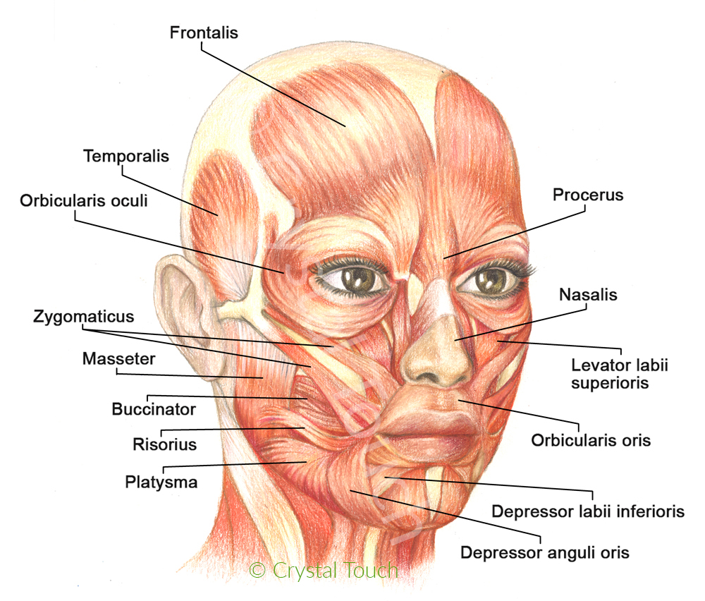
    Let’s take a closer look at our facial muscles, their names and functions. Please study facial muscles carefully. This will be important later, when we discuss the causes and the recovery from facial asymmetry and from synkinesis.


    Synergists and antagonists. Muscles that “help” each other to produce a certain movement are called synergists. Muscles that produce movements in opposite directions are called antagonists.


    Frontalis muscle lifts the eyebrows, makes horizontal forehead wrinkles when we are surprised.


    Orbicularis oculi – circular muscle of the eye. Closes the eyelids, squints the eye. These two muscles are antagonists. Lift and hold your eyebrow with your finger and then try to squint your eyes. Difficult, isn’t it?


    Procerus is a frown muscle. Pulls medial sides of eyebrows down and together.


    Corrugator superclii – pulls eyebrows together.


    Zygomatic muscles (major and minor) move the mouth corners up- and outward when we smile.


    Risorius – “smile” muscle. Pulls mouth corners laterally (outward) and forms dimples in the cheeks. This muscle is not always active in all people.


    Orbicularis oris (circular muscle of the mouth) puckers lips and brings mouth corners towards the middle line.


    Depressor anguli oris pulls mouth corners downward.


    Levator labii superioris and depressor labii inferioris pull the upper and lower lips up- and down respectively when we grin.


    Mentalis (chin muscle) pulls up the chin as we express disappointment, doubt and some other negative emotions.


    Platysma is a surface muscle of the neck. Platysma is engaged in the expressions of fear, disgust and some other negative emotions.


    The “crooked”, asymmetrical smile after Bell’s palsy happens when depressor anguli oris muscle on the affected side is unnecessarily activated together with zygomatic muscles (its antagonists) while Bell’s palsy sufferer wants to produce a smile. In this “fight” the former muscle often wins over the latter, simply because it is larger and stronger. Result: mouth corner on the affected side ”looks down” instead of “up”. Quite often several small dimples may appear on that side of the chin, leading to a further disfigurement of facial expressions.


    Facial Synkinesis is another common complication of long-standing Bell’s palsy. There are several opinions in the medical world about the causes of synkinesis. The most popular (and still controversial) is the theory of aberrant regeneration or simply said, re-connection of the regenerating branches of the facial nerve to the “wrong” muscles. As a conclusion of this theory, Synkinesis is considered to be an irreversible complication of Bell’s palsy.


    The results of an extensive scientific research at Crystal Touch clinic have demonstrated that with VERY few exceptions, facial synkinesis after long-standing Bell’s palsy have different origins. The actual cause of synkinesis is closely related to the higher cerebral activity. The recovery results of our numerous patients after 5, 10 and even 25 years since the onset of Bell’s palsy, prove that SYNKINESIS IS REVERSIBLE.


    2 2


    Crystal Touch Bell’s palsy clinic presented the results of our scientific research during the World Congress for NeuroRehabilitation in Philadelphia, USA (10-13 May 2016). Read more…


    – Alex PashovAlex

    Crystal Touch Bell’s palsy clinic


    Bell's Palsy Video Course


    Our Bell’s palsy educational video course provides a clear and elaborate knowledge about Bell’s palsy, how it affects the facial nerve and facial muscles, as well as why and how various residuals and complications such as synkinesis, contractures, facial asymmetry and pains are formed. We have made these videos to help explain complex anatomical explanations with the help of visual animations.


    (Currently available in English language with English and Portuguese subtitles)


    Learn More


    News about Bell's palsy and the clinic


    Subscribe if you'd like to receive our news and updates about Bell's palsy research, the clinic's progress and achievements, news about our seminars, workshops and other helpful developments.


     Click here to subsribe


    (繼續閱讀...)
    文章標籤

    金台診所附設醫美 發表在 痞客邦 留言(0) 人氣(177)

    • 個人分類:
    ▲top
    • 9月 18 週三 201912:43
    • 公告:金台診所臉書被封閉.造成不便敬請原諒

    公告:金台診所臉書被封閉.造成不便敬請原諒.如有需要請轉至吳聖臨臉書加好友謝謝.
    (繼續閱讀...)
    文章標籤

    金台診所附設醫美 發表在 痞客邦 留言(0) 人氣(118)

    • 個人分類:醫學美容
    ▲top
    • 8月 29 週四 201914:22
    • Anatomy and physiology of the eyelids


    Anatomy and physiology of the eyelids


    Dr.Reda Gomah El Garia by Dr.Reda Gomah El Garia

     
     February 26, 2018

     
    in Ophthalmology


     0


    (繼續閱讀...)
    文章標籤

    金台診所附設醫美 發表在 痞客邦 留言(0) 人氣(14)

    • 個人分類:醫學美容
    ▲top
    • 8月 29 週四 201910:06
    • Yellow White Retinal Lesion Differential Diagnosis

    Yellow White Retinal Lesion Differential Diagnosis


    Yellow White Retinal Lesion Differential Diagnosis


    BY OPHTHALMOLOGY NOTES POSTED ON JUNE 23, 2015 2:05PM


    (繼續閱讀...)
    文章標籤

    金台診所附設醫美 發表在 痞客邦 留言(0) 人氣(7)

    • 個人分類:醫學美容
    ▲top
    12...47»

    個人資訊

    金台診所附設醫美
    暱稱:
    金台診所附設醫美
    分類:
    醫療保健
    好友:
    累積中
    地區:

    熱門文章

    • (471)韓式4D隆鼻.發明人 – 李喜永博士
    • (118)公告:金台診所臉書被封閉.造成不便敬請原諒
    • (58)最新式下眼瞼筋膜整形術+中臉小拉皮術
    • (24)拉提≠拉皮!(各種拉皮&詳細分析).拉皮案例
    • (7)Yellow White Retinal Lesion Differential Diagnosis

    文章分類

    • 醫學美容 (318)
    • 未分類文章 (1)

    最新文章

    • Summary of VF defects
    • 什麼是黃斑部病變? 文自康健知識庫
    • Coronavirus disease 2019
    • Drug-induced uveitis
    • 十架七言-7個受傷的心得醫治的方法
    • Muscles of facial expressions and how they work
    • 公告:金台診所臉書被封閉.造成不便敬請原諒
    • Anatomy and physiology of the eyelids
    • Yellow White Retinal Lesion Differential Diagnosis
    • Macular Hole Surgery

    最新留言

    • [18/05/13] d946022 於文章「祝全天上天下的母親.母親節及天天快樂.H...」留言:
      m4QT0uy1:1大牌專賣,包包 、衣服 、手錶、鞋子,全...
    • [16/01/11] Josie Tsai 於文章「在新的一年,新的復興的突破要臨到,神正在...」發表了一則私密留言
    • [16/01/11] Josie 於文章「在新的一年,新的復興的突破要臨到,神正在...」發表了一則私密留言

    動態訂閱

    文章精選

    文章搜尋

    誰來我家

    參觀人氣

    • 本日人氣:
    • 累積人氣: