Pseudoexfoliation syndrome facts ( just now in my clinic )
Pseudoexfoliation syndrome facts ( just now in my clinic )
• important cause of secondary open-angle glaucoma.
• Although the appearance of dandruff-like material is characteristic of this condition,the earliest sign is the deposition of pigment in the

• Pupil dilatation is poor and cataract extractionmay be difficult.
• Complications of cataract surgeryare also increased due to weakened zonules, which can lead to zonulysis and vitreous loss.
• 50% of patients, chronic open angle glaucoma occurs. lntraocular pressures are often unstable and can lead to severe glaucoma. Therefore, any evidence of raised intraocular pressure should be treated early.
• Open angle most common due to block of trabecular meshwork by pigments and exfoliating material
• Closed angle in two ways
Phacomorphic glaucoma in neglected cases
Anterior displaced iris-lens diaphragm due to weakened zonules ( pupillary block)
Pseudoexfoliation syndrome facts PowerPoint presentations :
Pseudoexfoliation syndrome
1. Pseudoexfoliation Syndrome Presenter: Dr. Gloria George Moderator: Dr. Ajay R. Kamath
2. Introduction…  Pseudoexfoliation syndrome (PXF) or exfoliation syndrome is the most common identifiable cause of open angle glaucoma When eye with PXF develops secondary open-angle glaucoma pseudoexfoliation glaucoma (PXG) Systemic disorder with important eye manifestations- open and closed- angle glaucoma, cataract with zonular instability. Also associated with increased systemic risk of cardiovascular disorders
3. Epidemiology and Genetics… More common in older age groups- late 60’s and early 70’s U/L or B/L- most U/L cases become B/L over 20 years 40% of patients with PXF develop PXG More common in women, but men at higher risk for glaucoma Geographically- Scandinavian countries, Europe, UK, Middle East and South East Asia High risk conferred by mutations in the LOXL1 gene at locus 15q22, coding for elastic fibre components of the ECM
4. Etiology and Pathogenesis… Grey-white fibrillary extracellular material composed of a protein core surrounded by GAG’s is produced by abnormal BM of ageing epithelial cells in the trabeculum, equatorial lens capsule, iris and ciliary body Deposited on the anterior lens capsule, zonules, ciliary body, iris, trabeculum, anterior vitreous face and conjunctiva
5. Inherited microfibrillopathy ultrastructural appearance of random 10 to 12 nm fibrils, arranged in a fibrillogranular matrix or coiled as spirals The material behaves like a sticky “Christmas tree” type of protein, aggregating a large number of elastic tissue and basement membrane proteins Polymorphisms in the coding of LOXL1 gene LOXL1 enzyme- essential for formation of elastin fibres
6. Current concept in the pathogenesis of PXS
7. Ocular and Systemic Sources Many cell types- lens capsule epithelium, iris epithelium, vascular endothelium, corneal endothelium, and Schlemm’s canal endothelium, conjunctiva Other extrabulbar sites- extraocular muscles, orbital septa, posterior ciliary arteries, vortex veins, and central retinal vessels PEX material demonstrated in lung, heart, liver, gallbladder, skin, kidney, and cerebral meninges Associated with a number of vascular disorders, hearing loss and Alzheimer disease.
8. Clinical and Pathological changes… Corneal changes (pseudoexfoliation endotheliopathy) Flakes of pseudoexfoliation material and pigment accumulation on the corneal endothelium (diffuse or as a vertical spindle similar to the Krukenberg spindle) Specular microscopy of the corneal endothelium -significantly lower than normal cell density and changes in cell size and shape.
9. Ultrastructural studies have revealed clumps of pseudoexfoliation material adhering to the corneal endothelium and incorporated into the posterior Descemet’s membrane.
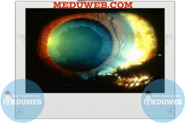
10. Iris changes (pseudoexfoliation iridopathy) Pseudoexfoliation material seen as white flecks on the pupillary margin of the iris, with loss of pigment at the pupillary ruff
11. Iris transillumination typically reveals a moth-eaten pattern near the pupillary sphincter or diffuse mid-peripheral transillumination defects.
12. Light and scanning electron microscopy demonstrate pseudoexfoliation material on the posterior surface of the iris Fluorescein angiographic studies of the iris have revealed hypoperfusion, peripupillary leakage, and neovascularization Vascular abnormalities or abnormal extracellular matrix production causes iris hypoxia atrophy of the iris pigment epithelium, stroma, and muscle cells Blood aqueous barrier defect, pseudo-uveitis
Pseudoeexfoliative glaucoma
Pseudoexfoliation syndrome facts videos:
Pseudoexfoliation Syndrome Plus Amiodarone – YouTube
Pseudoexfoliation Syndrome

 留言列表
留言列表